62 year old female with chronic cough and hemoptysis
This is an online E log book to discuss our patient's de-identified health data shared after taking his/her/guardian's signed informed consent.
Here we discuss our individual patient's problems through series of inputs from available global online community of experts with an aim to solve those patient's clinical problems with collective current best evidence based inputs.
This E log book also reflects my patient-centered online learning portfolio and your valuable inputs on the comment box is welcome.
Here is a case i have seen:
On 09th march 2021
A 62 year old female came to OPD with chief complaints of
cough with expectoration,
SOB since one month,
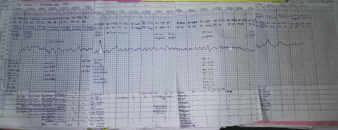
fever since one month,
vomitings and loose stools since 4 days
patient was apparently asymptomatic 20 years back later she had intermittent cough ( once in a month )which is relieved on taking medication but one month ago she had fever which was high grade with chills associated with low backache and SOB associated with cough with expectoration which was insidious in onset for this she went to hospital where she diagnosed with anaemia managed conservatively and she is on ceftriaxone. Symptoms aggravated more since 4 days , blood in sputum and wheeze present.
H/o vomitings since 4 days which are 4-6 episodes/ day,food and water as content,non bile stained, non blood stained.
H/o loose stools since 4 days 5-6 episodes/day.
H/o loss of appetite present.
Significant weight loss present
No h/o burning micturition,melaena, headache.
No history of hypertension, diabetes mellitus, epilepsy,thyroid disorders, asthma, tuberculosis.
No past surgical history and blood transfusions.
Family history -
- Her brother had similar complaints, died 25 years ago.
- Her sister has similar complaints chronic cough and SOB takes inhalers.
Menstrual and marital history-
Attained menarche 13 years of age and married at 14 years.
Menstrual cycles are regular 4/30,
Attained menopause by 45 years of age.
No known drug and allergies.
General examination-
PT is conscious, orientated to time, place, person, cooperative.
Thin built and Malnourished.
Skin fold thickness : 4mm.
HT-158 cm
Wt- 33 kg
BMI-13.25
PALLOR +, koilonychia +, clubbing +( fluctuation +)
No signs of icterus,cyanosis, generalised lymphadenopathy,paedal edema.
Vitals-
Temperature-99.3F
PR- 105 Bpm
RR- 28 cpm
Bp- 100/60 mm of Hg
Head to toe Examination:
Temporal wasting present.
Shiny, bald ,bulky, red tongue.
Greyish white patch is seen over soft palate.
Muscle wasting present: temporalis, deltoid.
Ichthyotic skin present on upper limbs and lower limbs.
?hard, mobile,2.5cm left supraclavicular lymph node present.
Lower thoracic and lumbar kyphoscoliosis present.
Lower limb : significant muscle wasting present.
Saddle nose deformity +
Respiratory examination:
Inspection-
Oral cavity- poor oral hygiene
UR-:12- 45678. UL:12345678.
LR:123--- 78. LL :12---- 78.
Cervical Trachea appears to be central.
Trail’s sign+.
Drooping of shoulder , on left side
Broadbent’s sign +:systolic retraction in 3rd and 4th ICS.
Kyphosis seen with medial border prominence of Scapula
Dilated veins seen over neck, right upper anterior aspect and left hemithorax.
Barrel shaped chest.
Suprasternal pulsations present
visible pulsations present in left mid clavicular line below the nipple( 4cm).
Epigastric pulsations +.
Posteriorly left side lower thoracic region - ?aortic pulsations present.
Abdominothoracic type respiration
Resp. movements Right. Left.
Upper zone ✓ Decreased
middle zone ✓ Decreased
Lower zone ✓ ✓
Accessory muscle usage present.
1.SCM
2.Scalenus
Palpation-
Trachea- Mediastinal tracheal shift to right
no local rise of Temperature and tenderness.
Left side over crowding of ribs +.
Apex beat felt over left to left mid axillary line in 5th intercostal space (4cm).
Anteroposterior diameter(APD)- 24 cm.
Transfers diameter(TD)-24cm.(APD/TD: 1/1).
Resp. Movements. Right Left.
Anterior:
Upper zone. N Decreased
middle zone N Decreased
Lower zone N Decreased
Posterior:
Suprascapular. N Decreased
Interscapular. N Decreased
Infrascapular. N Decreased
Percussion-
Direct : resonant over clavicular, sternum.
Indirect :
Anterior. Right Left.
Supraclavicular. Resonant. Dull
Infraclavicular. Resonant Flat
Supra mammary Resonant Flat
Mammary. Resonant Flat
Inframammary. Dull. Flat
Axillary. Resonant. Dull
Infraaxillary. Dull. Dull
Posterior:. Right. Left.
Suprascapular. Resonant. Dull
Interscapular. Resonant stony dull
Infrascapular. Resonant. Flat
Auscultation- decreased air entry in both the lung areas. bilateral coarse Crepitations heard in both the lung areas.
Aegophony and bronchophony in
Right. Left
Supraclavicular. ✓ tubulobronchi.
Infraclavicular. ✓ tubulobronchi
Supra mammary A&B. tubulobronchi
Mammary. A&B tubulobronchi
Inframammary. ✓. tubulobronchi
Axillary. ✓ ✓
Infraaxillary. ✓. ✓
Suprascapular. ✓. Tubulobronchi
Interscapular. ✓. Tubulobronchi
Infrascapular. ✓. Tubulobronchi
Per abdomen-
Distended abdomen, everted umbilicus present. Distended abdominal veins.
Shifting dullness present.mild spleenomegaly.
Bowel sounds heard.
? Portal hypertension
CVS- S1,S2 heard.
CNS- No focal neuronal deficits
Reflex's Right. Left.
Jaw jerk. +. +
Schimizu +. +
Biceps. +++. +++
Triceps. +++. +++
Supinator. +++. +++
Finger flexor. +++. +++
Knee. +++. +++
Ankle. +++. +++
Plantar. +++. +++
Investigations-
ABG-
PH- 7.24
PCO2- 32 mm hg
Po2- 79.3 mm hg
Hco3- 13.2 mmol/L
2D - Echo
2D - Echo
Moderate Tricuspid Regurgitation(TR) + with Pulmonary artery hypertension
Mild Mitral Regurgitation+/Aortic Regurgitation+.
Left Anterior Descending hypokinesia, Right Coronary Artery Left circumflex hypokinetic ,no Aortic Stenosis /Mitral Stenosis.
Moderate Left Ventricular dysfunction+
Diastolic dysfunction +, No Pulmonary Embolism.
USG-
B/L grade 1 Renal Parenchymal Disease changes present.
BT- 2 Min
CT- 4 Min
PR- 15
INR- 1.11
APTT- 30
Hb- 13.4
Anti HCV- Negative
HBsAg-negative
HIV1/2- negative
DENGUE- negative
Chest xray-


